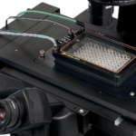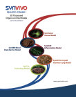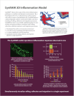
The SynRAM™ 3D Inflammation Model from SynVivo was developed to study the entire inflammation pathway in a dynamic environment. The SynVivo platform delivers a physiologically realistic model (including flow and shear) and enables real-time tracking of rolling, adhesion, and migration processes. The model recreates a histological slice of co-cultured tissue and/or tumor cells with a lumen of endothelial cells. It has been successfully validated against in vivo studies showing excellent correlation with rolling velocities, adhesion patterns, and migratory processes (Lamberti et al 2014, Soroush et al 2016).
The SynRAM 3D inflammation model provides a realistic testing environment including:
- Physiological shear stress within a microvascular environment
- In vivo like vascular morphology with fully enclosed lumen
- Co-culture capability for cell-cell interactions
- Quantitative real-time rolling, adhesion, and migration data from a single experiment
Leukocyte Endothelium interactions on-a-chip
SynRAM enables assessment of cellular interactions comprising of rolling, adhesion, and migration through multiple cellular layers in one experiment, in real-time, and represents data closely correlated with in vivo results.
SynRAM’s innovative design overcomes the current limitations inherent in flow chambers or Transwell chamber based assays. Current flow chamber designs are oversimplified, lack the scale and geometry of the microenvironment, and cannot model transmigration. Similarly, Transwell chambers do not account for fluid shear and size/topology observed in vivo. In addition, endpoint measurements of migration are not reproducible in a Transwell chamber and do not provide real-time visualization.
SynVivo’s proprietary chip designs range from complex in vivo derived microvascular networks (obtained from digitized images) to simplified idealized networks. Complex in vivo networks produce realistic cellular makeup and vascular morphology resulting in varying shear and flow conditions. Simplified idealized networks are designed to reproduce cellular makeup, constant shear, and flow conditions.
Examples of Models Functionalized in SynRAM Devices
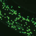
Real-time visualization of rolling, adhesion and migration
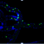
Real-time tracking of Single-cell migration
Leukocyte endothelium interactions
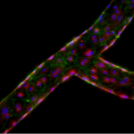
Vessel on a Chip with endothelial cells
PRODUCT PURCHASING OPTIONS
Chips: Depending on your specific research applications you can select from IMN2 radial SMN2 microvascular network chip configurations.
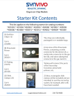
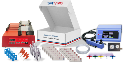
Kits: All the basic components required to run SynRAM assays can be purchased in a kit format. Two Kit formats are available.
Starter Kit: Select this for your first-time purchase
- 10 SynRAM chips (Choice of IMN2 radial or SMN2 microvascular network chips)
- Accessories including tubing, clamps, needles, and syringes
- Pneumatic priming device (required for priming tubing to remove air)
Assay Kit: Select this kit format if you have previously purchased the pneumatic priming device
- 10 SynRAM chips (Choice of IMN2 radial or SMN2 microvascular network chips)
- Accessories including tubing, clamps, needles, and syringes
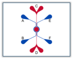
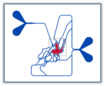
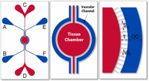
Schematics of the devices used to develop SynRAM inflammation models. Apical chamber (outer channels) are for culture of vascular (endothelial cells) while basolateral chamber (central chamber) are for culture of tissue cells. Porous architecture enables communication between the vascular and tissue cells.
Idealized Co-Culture Network Chips (IMN2 radial)
IMN2 radial Chip: (200μm Outer channel, 1.8mm tissue chamber, Pillars, 8um gap, 50μ travel (space between channels, 100μm depth (height). Gap, 50um Travel—–3/100 um height of pillars. Chip Cat#: 102008.
Starter Kit
$1,700Add to cart
Assay Kit
$1,500Add to cart
SMN2 microvascular network Co-Culture Chips
SMN2 microvascular Chip: 3 um Height Barrier: 10um Dia-50um Separation, 100 um Depth. Cat#: 105001.
Starter Kit
$2,100Add to cart
Assay Kit
$1,700Add to cart
Bioinspired Microfluidic Assay for In Vitro Modeling of Leukocyte–Endothelium Interactions
Authors: G. Lamberti, B. Prabhakarpandian, C. Garson, A. Smith, K. Pant, B. Wang, and M.F. Kiani. Anal.
Chem., 2014, 86 (16), pp 8344–8351 DOI:10.1021/ac5018716
SynRAM microfluidic chips comprising of realistic microvascular networks were used to understand the role of classical inhibitors of individual steps of the leukocyte adhesion cascade. Experimental results matched very well with in vivo data highlighting the unique ability of the platform for real-time analysis of these dynamic events in a morphologically realistic environment (Lamberti et al 2014).
Rolling, adhesion, and migration of neutrophils in bMFA; migration of neutrophils (labeled with fluorescent dye) into the tissue compartment of bMFA after 120 min of continuous flow. (1 and 2) Solid arrows in the top right panels show a rolling neutrophil which (3) becomes adherent; dotted arrows in the top right panels show firmly adherent neutrophils. A neutrophil migrating from a vascular channel through the barrier into the tissue compartment over time (bottom right).
Neutrophil rolling using SynRAM microfluidic chips is similar to leukocyte rolling in vivo; Box and whisker plots summarizes the comparison of leukocytes rolling velocity measured in vivo and in SynRAM chips and shows no significant difference (p=0.758; Mann-Whitney Rank Sum Test). The “+” marked in the box indicates the mean.
Neutrophil adhesion in SynRAM microfluidic chips is similar to leukocyte adhesion in vivo; Distribution of the number of adhered leukocytes and neutrophils as a function of distance from the nearest bifurcation in vivo in mouse cremaster muscle model and in vitro in microfluidic chips, respectively. Both histograms are skewed to the left indicating that leukocytes and neutrophils preferentially adhere near bifurcations with the peak occurring at one vessel or channel diameter from the nearest bifurcation.
Investigation of the Effect of Blocking of Specific Steps of the Inflammation Pathway using Monoclonal Antibodies
Antibody blocking of specific steps in the adhesion/migration cascade downregulates other steps of the cascade; Monoclonal antibodies against E-selectin (aE-selectin), ICAM-1(aICAM-1), and PI3K (wortmannin) significantly reduced the number of rolling, adhering, and migrating neutrophils in SynRAM microfluidic devices.
Antibody blocking of specific steps in the adhesion/migration cascade downregulates other steps of the cascade; monoclonal antibodies against E-selectin (aE-selectin), ICAM-1(aICAM-1), and PI3K (wortmannin) significantly reduced the number of rolling, adhering, and migrating neutrophils in bMFA. The numbers represent the percentage of activity after treating cells with the respective blockers in comparison to their corresponding control values (mean ± SEM; N = 3).
Elucidation of the Mechanism of Protein Kinase C delta (PKCδ) in Sepsis Related Inflammation Response
A Novel Microfluidic Assay Reveals a Key Role for Protein Kinase C δ in Regulating Human Neutrophil-Endothelium Interaction
Authors: Soroush F, Zhang T, King DJ, Tang Y, Deosarkar S, Prabhakarpandian B, Kilpatrick LE, Kiani MF.
J Leukoc Biol November 2016 100:1027–1035.
The SynRAM model was used to identify the underlying mechanism of Protein Kinase C delta (PKCδ) dependent neutrophil-endothelium interactions. These interactions have been found to play a significant role in the inflammatory response. They found that PKC𝛿 was a critical regulator of human neutrophil adhesion and migration through human endothelial cells during inflammation. This was validated by testing physiological fluid flow conditions of the entire inflammation process comprised of rolling, adhesion, and migration in real-time.
PKCδ inhibitor significantly reduces migration of neutrophils from the vascular channels, across the inflammed endothelium (treated with TNF-α for 4 or 24 hour), into the tissue compartment in response to fMLP mediated signaling compared to untreated controls.
Immunohistochemical detection of myeloperoxidase (MPO) in representative lung tissue sections from 24 h post surgery. Few MPO-positive cells in Sham surgery. Sepsis induces the infiltration of numerous MPO-positive cells throughout the lung parenchyma. PKCδ-TAT Inhibitor significantly reduces sepsis-induced, MPO-positive cell numbers in the lung indicating decreased neutrophil migration.
Chemotaxis and Migration
Cells are incubated in the vascular channel and chemoattractant is injected into the tissue chamber or the second vascular channel (if using the idealized network). Migration of immune cells across the engineered porous region into the tissue chamber is observed in real-time. The assay can be performed under static or flow conditions.
Vascular (endothelial cells) Mediated Rolling, Adhesion and Migration Assay
Endothelial cells cultured in the vascular channel are activated with biological or chemical agents to produce an inflammatory response followed by real-time tracking of rolling, adhesion and migration of immune cells across the endothelium into the tissue chamber.
Vascular and Tissue Co-culture Mediated Rolling, Adhesion and Migration Assay
Endothelial cells are cultured in vascular channel while tissue chamber houses smooth muscle cells, epithelial cells, fibroblast, or organ-specific cells (e.g. astrocytes, hepatocytes). In response to a cellular signaling and inflammation activation, rolling, adhesion, and migration across the endothelium can be observed and quantified in real-time.
Rolling, adhesion and migration of WBCs across the inflamed vascular-tissue interface.
Vessel on-a-chip applications can be functionalized using either of the SynVivo devices for drug delivery, vascular injury, endothelium-immune cell interactions or other applications
HUVEC endothelial cells under flow
Primary rat brain endothelial cells underflow
hCMEC/D3 brain endothelial cells under flow
Applications and Endpoints using the SynRAM model:
Models Available:
- Monoculture using primary endothelial cells/cell line
- Co-Culture with stromal/tissue cells
Assays:
- Immune cells (primary, cell lines) rolling, adhesion and migration across the endothelium
- Inflammation-induced vascular permeability
- Drug-induced vascular injury
- Inflammation-induced biomarker analysis
- Therapeutic screening
Sample Endpoints
Quantitation of immune cell interactions with the endothelium. Vascular permeability using fluorescent-tagged molecule, ROS, biomarkers screening using immunoassays, genomic, proteomic, or metabolomic analysis.
Contact us for a Services quote!
Distinct functional neutrophil phenotypes in sepsis patients correlate with disease severity
Authors:
Neutrophil‐Endothelial Interactions of Murine Cells is Not a Good Predictor of their Interactions in Human Cells
Authors: Fariborz Soroush, Yuan Tang, Omar Mustafa, Shuang Sun, Qingliang Yang, Laurie E. Kilpatrick, Mohammad F. Kiani
(2020) FASEB Volume 34, Issue 2: 2691-2702
The Role of Tyrosine Phosphorylation of Protein Kinase C Delta in Infection and Inflammation
Authors: Yang Q, Langston JC, Tang Y, Kiani MF, Kilpatrick LE.
Int J Mol Sci. 2019 Mar 26;20(6)
PKCδ Inhibition as a Novel Medical Countermeasure for Radiation-Induced Vascular Damage
Authors: Fariborz Soroush, Yuan Tang, Hasan M. Zaidi, Joel B. Sheffield, Laurie E. Kilpatrick, and Mohammad F. Kiani.
The FASEB Journal Vol. 32, No. 12. (2018)
A novel microfluidic assay reveals a key role for protein kinase C δ in regulating human neutrophil-endothelium interaction
Authors: Soroush F, Zhang T, King DJ, Tang Y, Deosarkar S, Prabhakarpandian B, Kilpatrick LE, Kiani MF.
J Leukoc Biol November 2016 100:1027–1035.
Adhesion Patterns in the Microvasculature are Dependent on Bifurcation Angle
Authors: G. Lamberti, F. Soroush, A. Smith, M. Kiani, B. Prabhakarpandian, K. Pant.
Microvascular Res., 2015, 99, pp 19-25
Bioinspired Microfluidic Assay for In Vitro Modeling of Leukocyte–Endothelium Interactions
Authors: G. Lamberti, B. Prabhakarpandian, C. Garson, A. Smith, K. Pant, B. Wang, and M.F. Kiani. Anal.
Chem., 2014, 86 (16), pp 8344–8351
Adhesive Interaction of Functionalized Particles and Endothelium in Idealized Microvascular Networks
Authors: G. Lamberti, Y. Tang, B. Prabhakarpandian, Y. Wang, K. Pant, M.F, Kiani, B. Wang.
Microvascular Res. 2013 (89) pp 107-114
Preferential Adhesion of Leukocytes Near Bifurcations is Endothelium-Independent
Authors: Tousi N, Wang B, Pant K, Kiani MF, Prabhakarpandian B.
Microvasc Res. 2010 Dec;80(3):384-8
SynRAM 3D Model – Starter Kits
Includes consumables (10 chips, 100ft tubing, 25 slide clamps, 50 blunt tip needles and 50 1 ml syringes). Starter kits include the pneumatic priming device.
Note: Does not include other required consumables such as cells, media, and matrix. Laboratory equipment required includes incubators, inverted microscopes, and syringe pumps.
CAT# | Product Name | Price |
---|---|---|
401002 | ||
401004 |
SynRAM 3D Model – Assay Kits
Includes consumables (10 chips, 100ft tubing, 25 slide clamps, 50 blunt tip needles and 50 1 ml syringes).
Note: Does not include other required consumables such as cells, media, and matrix. Laboratory equipment required includes incubators, inverted microscopes, and syringe pumps.
CAT# | Product Name | Price |
---|---|---|
401001 | ||
401003 |
SynRAM 3D Model – Chips
Purchase single chips.
Note: Does not include other required consumables such as cells, media, and matrix. Laboratory equipment required includes incubators, inverted microscopes, and syringe pumps.
CAT# | Product Name | Price |
---|---|---|
102008-SR3 | ||
105001-SR3 |