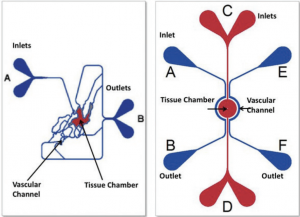
SynVivo’s proprietary microfluidic chips are capable of supporting a microvascular network that simulates the circulation inside any tissue with respect to flow, shear, and pressure. Novel co-culture protocols have been developed that establish true vascular monolayers in communication with tissue cells. Human cells grown in SynVivo chips retain biological phenotypes that are similar to cells found in the tissues. Leading researchers have validated that cells grown in SynVivo chips more accurately reflect the tissue cells found inside the body than do cells grown using conventional culture techniques.
The successful coupling of digitized tissue imaging with silicon etching technologies allows SynVivo to design and manufacture microfluidic chips that can be adapted for a variety of uses. All chip designs incorporate ports for the introduction of cells and reagents and for collecting effluent for analysis. They can accommodate virtually any analytical technology.
SynVivo has developed 3D Tissue and Organ-on-Chip models that accelerate real-time studies of cellular behavior, drug delivery, and drug discovery by providing a biologically realistic microenvironment that more accurately depicts in vivo reality. SynVivo models recreate complex in vivo microvasculature including scale, morphology, hemodynamic shear stress, and cellular interactions in an in vitro microfluidic chip environment. SynVivo 3D Tissue and Organ-on-Chip models enable real-time visualization and quantitation due to their side-by-side architecture.
